Introduction:
Breast cancer is a devastating disease that affects millions of individuals globally, making early detection and precise characterization critical for improving treatment outcomes. Over recent years, magnetic resonance imaging (MRI) has emerged as a powerful diagnostic tool in breast cancer management. This article aims to delve into the intricacies of breast MRI, including its fundamental principles, the MRI procedure for breast imaging, its role in breast cancer detection, and its use in characterizing breast cancer. Furthermore, we'll discuss the limitations of breast MRI and the exciting prospects it holds for the future of breast cancer diagnosis.
Part 1: Basics of Magnetic Resonance Imaging (MRI)
1.1. General Principles of MRI: Magnetic Resonance Imaging operates on the principle of nuclear magnetic resonance. It involves subjecting the body to strong magnetic fields and radio waves. In response to these forces, hydrogen protons in the body emit radiofrequency signals. The MRI machine captures these signals and uses them to construct detailed cross-sectional images of the breast tissue. MRI is renowned for its exquisite soft tissue contrast, making it particularly valuable for breast imaging.
1.2. Advantages of MRI: One of the key advantages of MRI, especially in breast imaging, is that it does not use ionizing radiation, which is a concern with mammography. This makes MRI a safe option for repeated examinations. Additionally, MRI's superior soft tissue contrast enables the detection of lesions that might not be visible on mammograms or ultrasounds, particularly in dense breast tissue.
Part 2: Breast MRI Procedure
2.1. Patient Preparation: Before undergoing a breast MRI, patients are typically instructed to fast for several hours to avoid any artifacts caused by food in the digestive system. Additionally, patients should remove any metallic objects, as these can interfere with the MRI's magnetic field. Patients are also asked about medical history, allergies, and medications to ensure a safe procedure.
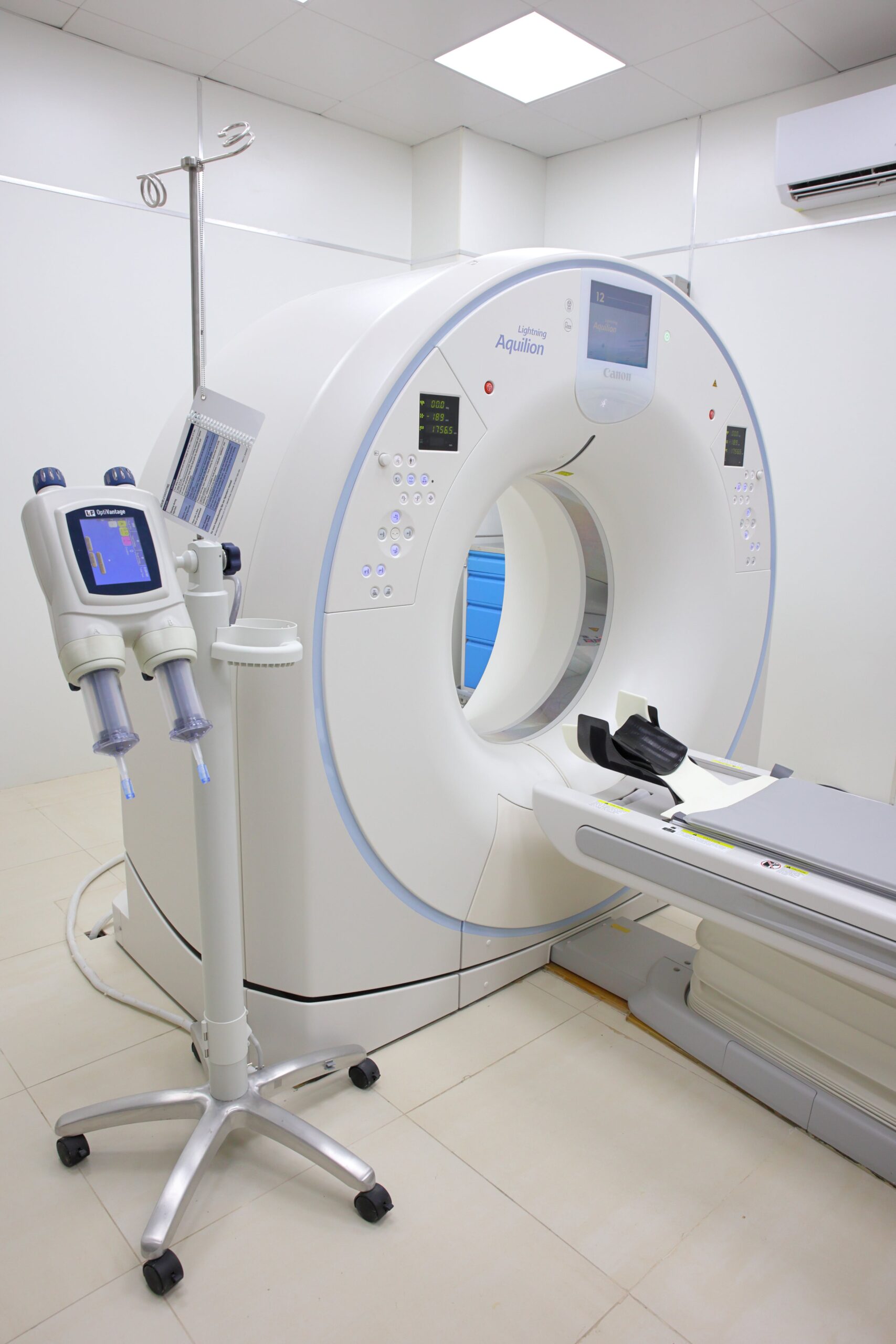
2.2. Conducting a Breast MRI: During the MRI procedure, the patient lies face down on the MRI table, and the breasts are placed in a specialized coil. The MRI machine emits radiofrequency signals, and a computer processes the signals to create detailed images of the breast tissue. The procedure is non-invasive and generally well-tolerated by patients.
Part 3: Utilizing MRI for Breast Cancer Detection
3.1. Role of Breast MRI in Secondary Screening: Breast MRI is frequently used as a secondary screening tool for individuals with suspicious findings on other breast imaging modalities, such as mammography or ultrasound. It provides additional information that can help confirm or rule out the presence of cancer. This is particularly valuable for high-risk individuals or those with dense breast tissue.
3.2. Role of Breast MRI in Primary Diagnosis: In certain cases, breast MRI serves as the primary diagnostic tool. This is especially true for high-risk individuals, such as those with a strong family history of breast cancer, or when mammography or ultrasound results are inconclusive. MRI can provide a more detailed assessment of breast abnormalities.
Part 4: Characterizing Breast Cancer with MRI
4.1. Distinguishing Tumor Types: MRI plays a pivotal role in differentiating various types of breast cancer. Different cancer types, like invasive ductal carcinoma, invasive lobular carcinoma, or ductal carcinoma in situ (DCIS), exhibit distinct imaging characteristics. This differentiation is crucial for treatment planning.
4.2. Evaluating Tumor Size and Staging: Accurate measurement of tumor size and staging is vital for developing an effective treatment plan. MRI provides precise measurements, which are crucial for determining the extent of the disease and guiding decisions regarding surgery, radiation therapy, and chemotherapy.
Part 5: Limitations and Future Perspectives
5.1. Limitations of Breast MRI: While breast MRI is a powerful diagnostic tool, it's not without limitations. These include the cost of the procedure, limited availability in some regions, and the potential for false-positive results, which can lead to unnecessary biopsies and anxiety for patients. It's important to carefully weigh these factors in clinical practice.
5.2. Future of Breast MRI in Breast Cancer Diagnosis: Ongoing research and technological advancements offer exciting prospects for the future of breast MRI. Innovations in contrast agents and image processing techniques are expected to improve the accuracy and efficiency of breast cancer diagnosis, making it an even more valuable tool for clinicians.
Conclusion:
Breast MRI has evolved into a valuable asset in the early detection and characterization of breast cancer. Its capacity to provide high-resolution images, assist in diagnosis, and differentiate between various tumor types contributes significantly to personalized and effective treatment strategies. While it has its limitations, ongoing research and technological advancements promise to enhance its role in breast cancer diagnosis, offering hope for better outcomes in the fight against this disease.
